Home
/ Paramecium Under Microscope : Paramecium Bursaria Light Microscopy Stock Video Clip K005 6668 Science Photo Library - Ciliated protozoans bog invertebrates protozoans.
Paramecium Under Microscope : Paramecium Bursaria Light Microscopy Stock Video Clip K005 6668 Science Photo Library - Ciliated protozoans bog invertebrates protozoans.
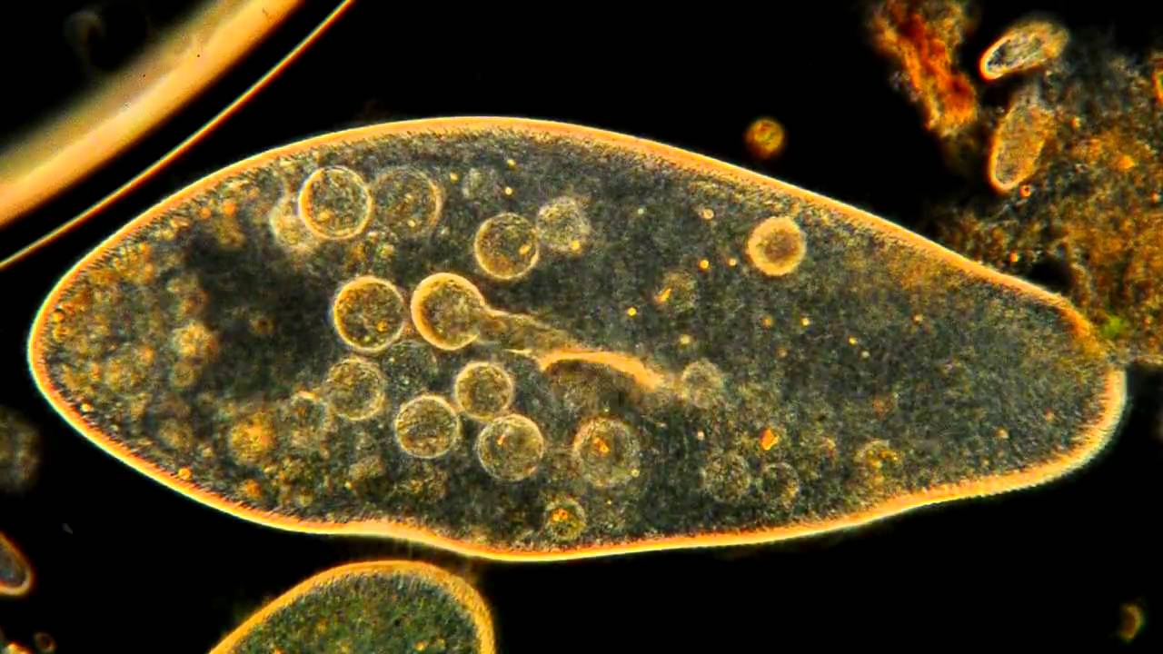
Paramecium Under Microscope : Paramecium Bursaria Light Microscopy Stock Video Clip K005 6668 Science Photo Library - Ciliated protozoans bog invertebrates protozoans.. Prepare the smear of the same and observe under compound microscope. They are easily maintained and cultured and paramecium caudatum (shown at the left) are characterized by a large macronucleus and a single compact micronucleus. Oral cilia and body cilia. Using a student biological microscope (also known as a compound microscope), you can grow some paramecium and watch as they swim around just like the video below. After a few days, place a drop of water from the jar on a slide and cover it with a cover slip.
Paramecium is a slipper shaped ciliate found in oxygenated aquatic environments feeding near vegetative matter. Paramecium under the microscope paramecium is a genus of the single cell ciliate protozoa and are found in freshwater marine areas and often in stagnant ponds. Paramecium are unique to microscopy because they were one of the first ciliates to be seen by microscopists in the late 17th century. What does paramecium look like under a microscope? Paramecia swim happily in ponds and streams throughout world.
Paramecium Bursaria Light Microscopy Stock Video Clip K005 6668 Science Photo Library from media.sciencephoto.com It is a eukaryote that has developed cellular organelles with a nucleus enclosed inside a nuclear membrane. Oral cilia and body cilia. Its last meal, like its first, was bacteria cells, neatly packed and ready for digestion. Under darkfield illumination at a magnification of 200x with a playing time of 21.0 seconds. It is a ciliated organism with cilia present throughout the body of the organism. Place a small drop of paramecium in the center of the microscope slide. What is the paramecium's displacement? As such, they are not plants, animal or fungi.
What does paramecium look like under a microscope?
Its size ranges from 170 to 290um or up to 300 to 350um. Then observe it under the microscope, starting at 40x. Then seal the lid and keep where it can get a lot of sunlight. Oral cilia are present on the surface of the oral groove. Cadatum is a microscopic, unicellular protozoan. The paramecium starts at the 65 mm mark and ends up at the 39 mm mark. They help collect food materials. They are easily maintained and cultured and paramecium caudatum (shown at the left) are characterized by a large macronucleus and a single compact micronucleus. Culturing pond life microscope talk. What does paramecium look like under a microscope? Its last meal, like its first, was bacteria cells, neatly packed and ready for digestion. Using a device called an electron microscope, we can look inside the food vacuoles and see what the paramecium has eaten. Place a small drop of paramecium in the center of the microscope slide.
in this video paramecium cilia movement under a microscope. Paramecium under microscope 40x e993 com. Its last meal, like its first, was bacteria cells, neatly packed and ready for digestion. Motion concepts problem 1.6 5 of 16 a review l constants periodic table part a logan observes a paramecium under a microscope. Paramecium is a slipper shaped ciliate found in oxygenated aquatic environments feeding near vegetative matter.
Amazing Microscopic Hd Video Paramecium Feeding Youtube from i.ytimg.com This video was captured using the zeiss primostar hd digital educational microscope. Express your answer with appropriate units. Its last meal, like its first, was bacteria cells, neatly packed and ready for digestion. Paramecium caudatum is a single celled ciliated protozoan that feeds on bacteria and other small microbes. Then observe it under the microscope, starting at 40x. Paramecium under the microscope paramecium is a genus of the single cell ciliate protozoa and are found in freshwater, marine areas, and often in stagnant ponds. Introduction to cell biology includes systems analysis cell theory characteristics of living things as well as compound light microscope use and structure and function of cell organelles. Paramecium can be found in lakes, ponds, rivers an.
Paramecium is a slipper shaped ciliate found in oxygenated aquatic environments feeding near vegetative matter.
Logan observes a paramecium under a microscope. Paramecium captured under the microscope at 400x. Paramecium caudatum is a single celled ciliated protozoan that feeds on bacteria and other small microbes. Paramecium under the microscope paramecium is a genus of the single cell ciliate protozoa and are found in freshwater marine areas and often in stagnant ponds. Paramecium under microscope 40x e993 com. Using a student biological microscope (also known as a compound microscope), you can grow some paramecium and watch as they swim around just like the video below. Its last meal, like its first, was bacteria cells, neatly packed and ready for digestion. Paramecium are unique to microscopy because they were one of the first ciliates to be seen by microscopists in the late 17th century. Paramecium sp bright field 400x clearly visible within the. in this video paramecium cilia movement under a microscope. Paramecium are grouped into a specialized category called ciliate because their cells contain small hair like structures on the exterior called cilia which the paramecium use for movement and to engulf their food. Mix thoroughly and carefully place a coverslip on the top. After a few days, place a drop of water from the jar on a slide and cover it with a cover slip.
Then observe it under the microscope, starting at 40x. Paramecium is a slipper shaped ciliate found in oxygenated aquatic environments feeding near vegetative matter. Cadatum is a microscopic, unicellular protozoan. With an electron microscope, we can examine the food vacuoles in high detail and see what the paramecium has eaten. The paramecium starts at the 65 mm mark and ends up at the 39 mm mark.
Electron Microscopy Of Paramecium Ciliata Sciencedirect from ars.els-cdn.com Motion concepts problem 1.6 5 of 16 a review l constants periodic table part a logan observes a paramecium under a microscope. In this video, you will see them move, feed, die, split, and clump together for no reason at all. Under darkfield illumination at a magnification of 200x with a playing time of 21.0 seconds. Mix thoroughly and carefully place a coverslip on the top. Oral cilia are present on the surface of the oral groove. Paramecium is a slipper shaped ciliate found in oxygenated aquatic environments feeding near vegetative matter. Today, we observe the paramecium! Cadatum is a microscopic, unicellular protozoan.
The eyepiece of the microscope has a horizontal scale marked in mm the paramecium starts at the 65 mm mark and ends up at the 39 mm mark what is the paramecium's displacement?
Paramecium under the microscope paramecium is a genus of the single cell ciliate protozoa and are found in freshwater, marine areas, and often in stagnant ponds. Paramecium caudatum is a single celled ciliated protozoan that feeds on bacteria and other small microbes. Oral cilia and body cilia. Only one species lives in marine waters. Paramecium at 400x magnification at microscope clip 87816927. Paramecium are grouped into a specialized category called ciliate because their cells contain small hair like structures on the exterior called cilia which the paramecium use for movement and to engulf their food. They are easily cultivated in the laboratory by allowing vegetable matter to stand in water for a few days. Animal conjugation of paramecium caudatum stock photo. The paramecium starts at the 65 mm mark and ends up at the 39 mm mark. Using a student biological microscope (also known as a compound microscope), you can grow some paramecium and watch as they swim around just like the video below. Today, we observe the paramecium! 400x magnification paramecium under microscope 400x. Paramecium can be found in lakes, ponds, rivers an.